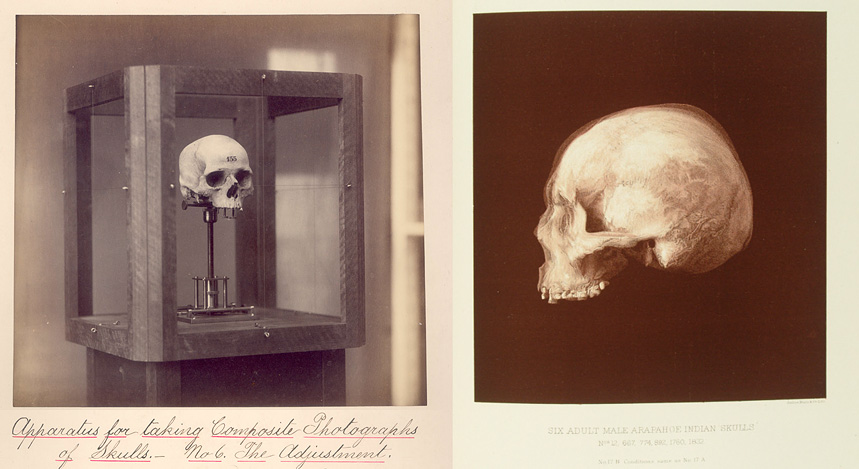
Ghostly photos from the Otis Archives depicting a novel circa-1885 piece of scientific analysis equipment: Apparatus for taking Composite Photographs of Skulls. Basically a wood and brass frame with a craniophore in the middle, the tool made it possible to position and align multiple skulls so composite photos could be taken accurately from the front, side, and rear views. The image on the right, for example, is a composite of five or six (or more?) separate skulls. From a contemporary anthropology journal describing the process:
Then the anterior frame and the lateral frame next to the window are lowered ; a black velvet background is hung on the posterior frame ; a large white cardboard is hung on the frame further from the window ; the brass-work is occluded with small velvet screens, and the picture is taken.
The photographs record composites of skulls from various Native American and Cook Island tribes (as seen in the archives of the Clark Institute), so I first thought that the measurements were sadly being undertaken for the sake of scientific racism, the darker side of physical anthropology, which was still in vogue in the 1880s.
That may be the case, but thankfully the full story is somewhat more complex: the inventor of the apparatus, Washington Matthews, an army surgeon-ethnographer-linguist, wrote extensively on the Siouan languages while stationed in the Dakotas, reportedly married and had a son with a woman from the Hidatsa tribe, was initiated into some aspects of the Navajo tribe, and also contributed substantially to the understanding and recording of the Navajo culture, which previously had been considered primitive by the Europeans:
Dr Matthews referred to Dr Leatherman’s account of the Navahoes as the one long accepted as authoritative. In it that writer has declared that they have no traditions nor poetry, and that their songs “were but a succession of grunts.” Dr. Matthews discovered that they had a multitude of legends, so numerous that he never hoped to collect them all: an elaborate religion, with symbolism and allegory, which might vie with that of the Greeks; numerous and formulated prayers and songs, not only multitudinous, but relating to all subjects, and composed for every circumstance of life. The songs are as full of poetic images and figures of speech as occur in English, and are handed down from father to son, from generation to generation.
Sorry for the icky photo, folks, just wanted to share a striking bit of anatomical illustration! This image led me down the rabbit hole of looking into the art of moulage, casting realistic wax models with “wounds” and other dermatological problems for use in medical training. Obviously a much better way of introducing a classroom full of doctors to diseases than wheeling in an actual smallpox patient. There’s a photo book on the subject called Diseases in Wax: The History of Medical Moulage that I might have to track down. At $180 on Amazon, though, I sure hope that the library here has it…
In the winter of 1511, Michelangelo entered the final stages of the Sistine Chapel project and painted 4 frescoes along the longitudinal apex of the vault, which completed a series of 9 central panels depicting scenes from the Book of Genesis. It is reported that Michelangelo concealed an image of the brain in the first of these last 4 panels, namely, the Creation of Adam. Here we present evidence that he concealed another neuronanatomic structure in the final panel of this series, the Separation of Light From Darkness, specifically a ventral view of the brainstem.
Could be pareidolia, but given Michelangelo’s breakthroughs in anatomical rendering and that God is depicted here with a rather non-standard bearded neck, who knows?
For those of you with eyes that aren’t easily categorized as simply “brown”, “blue”, or “hazel”: researchers have written up a new genetic model for human eye color phenotyping, published in PLoS Genetics.
We measured human eye color to hue and saturation values from high-resolution, digital, full-eye photographs of several thousand Dutch Europeans. This quantitative approach, which is extremely cost-effective, portable, and time efficient, revealed that human eye color varies along more dimensions than the one represented by the blue-green-brown categories studied previously. Our work represents the first genome-wide study of quantitative human eye color. We clearly identified 3 new loci, LYST, 17q25.3, TTC3/DSCR9, in contributing to the natural and subtle eye color variation along multiple dimensions, providing new leads towards a more detailed understanding of the genetic basis of human eye color.
I’ve actually wondered about this, as von Hagen’s popular (and assumed lucrative) Body Worlds exhibits have spawned many imitators of possibly questionable origins (one competing show is currently on view in a gallery inside our football stadium at the University of Texas). I guess that beyond the ethical and moral issues of consent and propriety, there’s also the business aspect: how enforceable is copyright when the primary medium is the deceased human body?
Animator Patrick Smith offers advice over on Scribble Junkies about drawing hands, an area of life drawing I still struggle with. He rails against both Preston Blair and Burne Hogarth’s popular treatises, and I’d have to agree with him there (I think the Hogarth “dynamic” books stunted my artistic abilities and understanding of anatomy by a few years, personally…).
One of the responses in the comments section rings true: “The drawings of hands you admire were probably drawn by people who looked at hands, not drawings of drawings of hands.”
A stylistically interesting medical drawing from the excellent Otis Historical Archives of the Walter Reed hospital: “Drawing. Life cycle of Balantidiasis”, part of a small set of other cartoons available on their Flickr stream. Gotta watch out for those protozoa.
Dreaming the Industrial Body. I’m not sure that I’m following the info that they’re trying to convey about the sun’s effects on our bodies, but it’s definitely an eye-catching image as far as medical literature goes. From Der mensch gesund und krank [The Healthy and Sick Human], menschenkunde 1940 … . Vol. 2, displayed as part of the National Library of Medicine’s Dream Anatomy exhibit. Speaking of which, check out their cool children’s art page!
Pioneer of medical instruments, photography, and cinema. Took some very interesting early photographs in his research of animal locomotion and physionomy, which led to his successor Muybridge’s famous collections of plates.